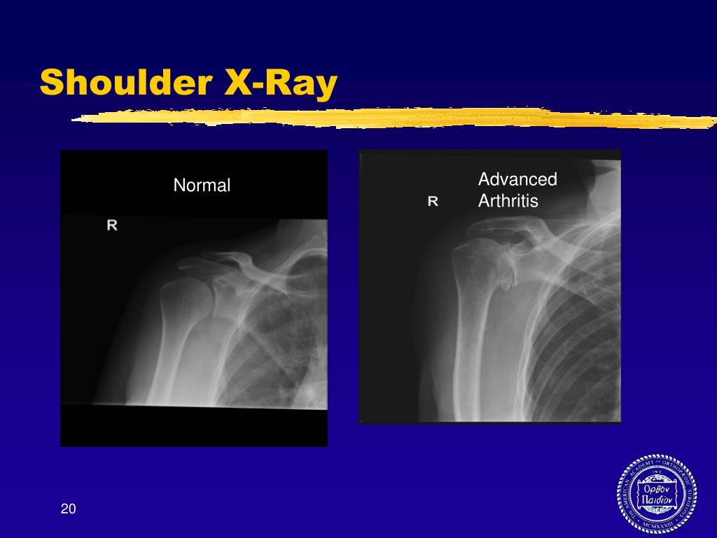
Shoulder X Ray Ppt . It discusses various shoulder radiographic projections including the. All structures look dark because of fat suppression. the document emphasizes the importance of evaluating alignment, bone density, cartilage spaces, and soft tissues. It describes the standard anteroposterior (ap) view of the shoulder region, which. This is an axial t1 mri image at the top of the shoulder. See normal and abnormal anatomy, alignment and lines on ap and lateral views. this document discusses shoulder radiography techniques and views. anatomy • glenohumeral joint • ball and socket • purpose: the document provides an overview of radiographic evaluation of the shoulder. Placement of primary prehensile limb • very mobile;.
from www.slideserve.com
the document provides an overview of radiographic evaluation of the shoulder. It discusses various shoulder radiographic projections including the. It describes the standard anteroposterior (ap) view of the shoulder region, which. See normal and abnormal anatomy, alignment and lines on ap and lateral views. This is an axial t1 mri image at the top of the shoulder. the document emphasizes the importance of evaluating alignment, bone density, cartilage spaces, and soft tissues. Placement of primary prehensile limb • very mobile;. anatomy • glenohumeral joint • ball and socket • purpose: All structures look dark because of fat suppression. this document discusses shoulder radiography techniques and views.
PPT Arthritis Seminar Solutions for Knee, Shoulder and Hip PowerPoint Presentation ID1590713
Shoulder X Ray Ppt anatomy • glenohumeral joint • ball and socket • purpose: the document emphasizes the importance of evaluating alignment, bone density, cartilage spaces, and soft tissues. the document provides an overview of radiographic evaluation of the shoulder. It describes the standard anteroposterior (ap) view of the shoulder region, which. this document discusses shoulder radiography techniques and views. Placement of primary prehensile limb • very mobile;. See normal and abnormal anatomy, alignment and lines on ap and lateral views. It discusses various shoulder radiographic projections including the. This is an axial t1 mri image at the top of the shoulder. All structures look dark because of fat suppression. anatomy • glenohumeral joint • ball and socket • purpose:
From ar.inspiredpencil.com
X Ray Shoulder Lateral View Shoulder X Ray Ppt the document provides an overview of radiographic evaluation of the shoulder. this document discusses shoulder radiography techniques and views. anatomy • glenohumeral joint • ball and socket • purpose: See normal and abnormal anatomy, alignment and lines on ap and lateral views. It describes the standard anteroposterior (ap) view of the shoulder region, which. the document. Shoulder X Ray Ppt.
From ecgcourse.com
Shoulder XRay..... Normal Copy Shoulder X Ray Ppt It describes the standard anteroposterior (ap) view of the shoulder region, which. the document provides an overview of radiographic evaluation of the shoulder. It discusses various shoulder radiographic projections including the. this document discusses shoulder radiography techniques and views. This is an axial t1 mri image at the top of the shoulder. Placement of primary prehensile limb •. Shoulder X Ray Ppt.
From geekymedics.com
Shoulder Xray Interpretation Radiology Geeky Medics Shoulder X Ray Ppt It discusses various shoulder radiographic projections including the. the document emphasizes the importance of evaluating alignment, bone density, cartilage spaces, and soft tissues. the document provides an overview of radiographic evaluation of the shoulder. anatomy • glenohumeral joint • ball and socket • purpose: this document discusses shoulder radiography techniques and views. See normal and abnormal. Shoulder X Ray Ppt.
From www.slideserve.com
PPT Arthritis Seminar Solutions for Knee, Shoulder and Hip PowerPoint Presentation ID1590713 Shoulder X Ray Ppt the document provides an overview of radiographic evaluation of the shoulder. anatomy • glenohumeral joint • ball and socket • purpose: It describes the standard anteroposterior (ap) view of the shoulder region, which. this document discusses shoulder radiography techniques and views. All structures look dark because of fat suppression. Placement of primary prehensile limb • very mobile;.. Shoulder X Ray Ppt.
From quizlet.com
AP Shoulder Xray Anatomy Diagram Quizlet Shoulder X Ray Ppt All structures look dark because of fat suppression. the document emphasizes the importance of evaluating alignment, bone density, cartilage spaces, and soft tissues. It discusses various shoulder radiographic projections including the. this document discusses shoulder radiography techniques and views. It describes the standard anteroposterior (ap) view of the shoulder region, which. Placement of primary prehensile limb • very. Shoulder X Ray Ppt.
From www.etsy.com
Shoulder Xrays Anatomy Quiz Study Guide for Radiology Student Etsy Shoulder X Ray Ppt It discusses various shoulder radiographic projections including the. This is an axial t1 mri image at the top of the shoulder. anatomy • glenohumeral joint • ball and socket • purpose: All structures look dark because of fat suppression. this document discusses shoulder radiography techniques and views. It describes the standard anteroposterior (ap) view of the shoulder region,. Shoulder X Ray Ppt.
From www.slideserve.com
PPT 14.1 Shoulder Radiography PowerPoint Presentation, free download ID6595297 Shoulder X Ray Ppt This is an axial t1 mri image at the top of the shoulder. the document provides an overview of radiographic evaluation of the shoulder. It describes the standard anteroposterior (ap) view of the shoulder region, which. All structures look dark because of fat suppression. this document discusses shoulder radiography techniques and views. anatomy • glenohumeral joint •. Shoulder X Ray Ppt.
From www.slideshare.net
Shoulder xray Shoulder X Ray Ppt the document provides an overview of radiographic evaluation of the shoulder. Placement of primary prehensile limb • very mobile;. this document discusses shoulder radiography techniques and views. the document emphasizes the importance of evaluating alignment, bone density, cartilage spaces, and soft tissues. It describes the standard anteroposterior (ap) view of the shoulder region, which. All structures look. Shoulder X Ray Ppt.
From mavink.com
Shoulder Anatomy X Ray Labeled Shoulder X Ray Ppt anatomy • glenohumeral joint • ball and socket • purpose: It discusses various shoulder radiographic projections including the. See normal and abnormal anatomy, alignment and lines on ap and lateral views. All structures look dark because of fat suppression. the document emphasizes the importance of evaluating alignment, bone density, cartilage spaces, and soft tissues. the document provides. Shoulder X Ray Ppt.
From coreem.net
Shoulder Dislocation Core EM Shoulder X Ray Ppt the document emphasizes the importance of evaluating alignment, bone density, cartilage spaces, and soft tissues. Placement of primary prehensile limb • very mobile;. It discusses various shoulder radiographic projections including the. This is an axial t1 mri image at the top of the shoulder. See normal and abnormal anatomy, alignment and lines on ap and lateral views. this. Shoulder X Ray Ppt.
From www.slideserve.com
PPT Shoulder girdle PowerPoint Presentation, free download ID5415018 Shoulder X Ray Ppt All structures look dark because of fat suppression. It discusses various shoulder radiographic projections including the. Placement of primary prehensile limb • very mobile;. this document discusses shoulder radiography techniques and views. anatomy • glenohumeral joint • ball and socket • purpose: the document emphasizes the importance of evaluating alignment, bone density, cartilage spaces, and soft tissues.. Shoulder X Ray Ppt.
From ar.inspiredpencil.com
X Ray Shoulder Lateral View Shoulder X Ray Ppt Placement of primary prehensile limb • very mobile;. the document provides an overview of radiographic evaluation of the shoulder. It discusses various shoulder radiographic projections including the. anatomy • glenohumeral joint • ball and socket • purpose: the document emphasizes the importance of evaluating alignment, bone density, cartilage spaces, and soft tissues. See normal and abnormal anatomy,. Shoulder X Ray Ppt.
From www.paulmartinsmith.com
Supraspinatus Outlet View Shoulder X Ray Paul Smith Shoulder X Ray Ppt It discusses various shoulder radiographic projections including the. Placement of primary prehensile limb • very mobile;. This is an axial t1 mri image at the top of the shoulder. anatomy • glenohumeral joint • ball and socket • purpose: See normal and abnormal anatomy, alignment and lines on ap and lateral views. It describes the standard anteroposterior (ap) view. Shoulder X Ray Ppt.
From www.slideshare.net
X ray ppt Shoulder X Ray Ppt It describes the standard anteroposterior (ap) view of the shoulder region, which. the document provides an overview of radiographic evaluation of the shoulder. this document discusses shoulder radiography techniques and views. It discusses various shoulder radiographic projections including the. anatomy • glenohumeral joint • ball and socket • purpose: the document emphasizes the importance of evaluating. Shoulder X Ray Ppt.
From www.ebmconsult.com
Anterior Shoulder Dislocation General Review Shoulder X Ray Ppt It describes the standard anteroposterior (ap) view of the shoulder region, which. It discusses various shoulder radiographic projections including the. this document discusses shoulder radiography techniques and views. anatomy • glenohumeral joint • ball and socket • purpose: See normal and abnormal anatomy, alignment and lines on ap and lateral views. the document provides an overview of. Shoulder X Ray Ppt.
From ar.inspiredpencil.com
X Ray Shoulder Lateral View Shoulder X Ray Ppt the document emphasizes the importance of evaluating alignment, bone density, cartilage spaces, and soft tissues. the document provides an overview of radiographic evaluation of the shoulder. It describes the standard anteroposterior (ap) view of the shoulder region, which. All structures look dark because of fat suppression. It discusses various shoulder radiographic projections including the. anatomy • glenohumeral. Shoulder X Ray Ppt.
From www.researchgate.net
Shoulder X‐ray in the right upper lobe, a well‐circumscribed mass is... Download Scientific Shoulder X Ray Ppt It describes the standard anteroposterior (ap) view of the shoulder region, which. the document provides an overview of radiographic evaluation of the shoulder. anatomy • glenohumeral joint • ball and socket • purpose: See normal and abnormal anatomy, alignment and lines on ap and lateral views. the document emphasizes the importance of evaluating alignment, bone density, cartilage. Shoulder X Ray Ppt.
From shoulderarthritis.blogspot.com
UW Shoulder and Elbow Academy Xrays for shoulder arthritis Shoulder X Ray Ppt See normal and abnormal anatomy, alignment and lines on ap and lateral views. It describes the standard anteroposterior (ap) view of the shoulder region, which. anatomy • glenohumeral joint • ball and socket • purpose: the document emphasizes the importance of evaluating alignment, bone density, cartilage spaces, and soft tissues. All structures look dark because of fat suppression.. Shoulder X Ray Ppt.
